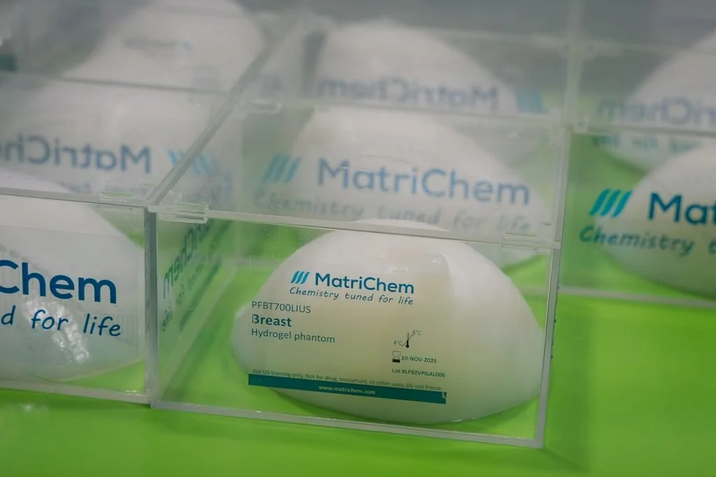
Our ultrasound breast phantom is designed for realistic sonography and biopsy training, mimicking the acoustic properties of human breast tissue and common lesions such as cysts, fibroadenomas, and cancers. Developed in partnership with The Bulgarian Breast Cancer Association, it supports high-quality, standardized education for radiologists and clinicians.
Key Features
Realistic ultrasound imaging with tissue-mimicking acoustic properties
Includes common lesions: cysts, fibroadenomas, and tumors (benign & malignant)
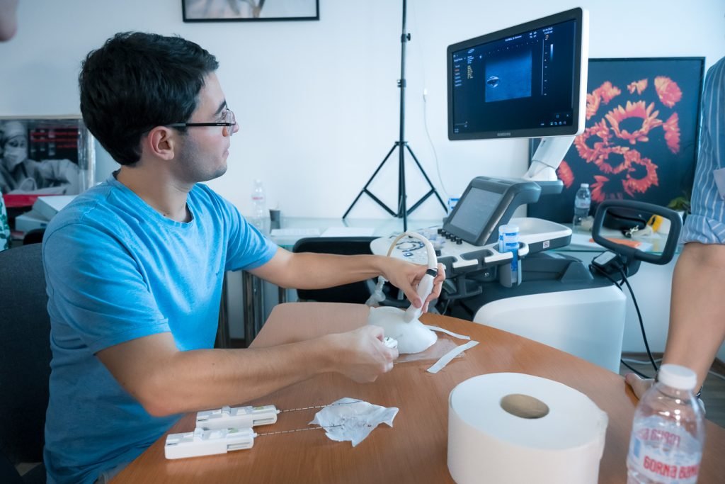
Suitable for ultrasound-guided biopsy training and needle insertion practice
Durable gel-based construction designed for repeated procedures
Available in multiple configurations depending on course size & training needs

Technical Specifications
Phantom material: Tissue-mimicking hydrogel
Acoustic properties:
Speed of sound: ~1520 m/s
Attenuation coefficient: ~0.30 dB/cm/MHz
Density: ~1.02 g/cm³
Lesion types included:
Anechoic oval cyst
Hypoechoic oval fibroadenoma
Irregular cancerous masses
Lesion sizes: 15–30 mm
Phantom dimensions: 140 mm × 140 mm × 70 mm
Compatible imaging modalities:
Ultrasound (5–15 MHz probes)
Biopsy capability:
Supports needle insertions
Self-healing
Durability:
Withstands ~50–100 needle passes
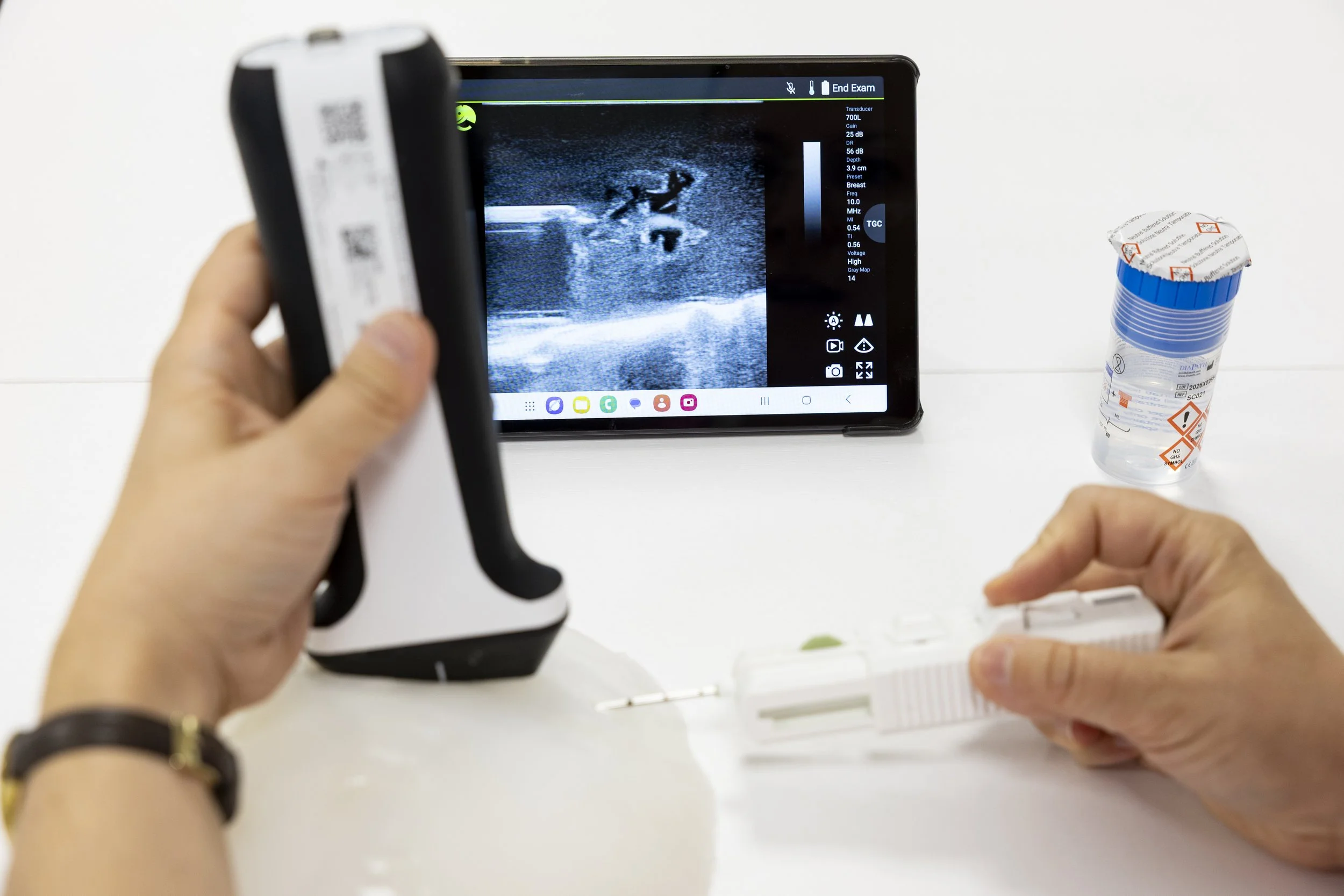
Ultrasound image of phantom with core needle

Ultrasound image of phantom with core needle
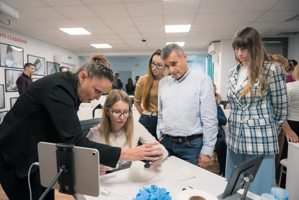
Benefits for Training & Education
The use of tissue-like phantoms is important for education of medical specialists for various reasons:
Realistic ultrasound simulation
The phantom provides tissue-mimicking acoustic properties that closely resemble human breast tissue, enabling trainees to practice probe handling, lesion localization, and depth assessment. Consistent lesion appearance across sessions allows learners to build confidence with reliable, repeatable images.
Safe and Effective Biopsy Training
Designed for ultrasound-guided interventions, the phantom allows trainees to perform needle insertion and targeting exercises without risk to patients. Internal structures respond realistically during biopsy practice, helping learners develop accurate hand–eye coordination and improve procedural proficiency.
Standardized Learning for Groups
Because every phantom contains the same lesion types and image characteristics, training programs can ensure consistent learning outcomes across large groups. This supports objective skills assessment, efficient teaching in workshops or courses, and enables repeated practice in a controlled environment.

Types of Breast Phantom Lesions
Cyst
Anechoic, fluid-filled cysts appear as black circular structures under ultrasound. Our phantom reproduces their distinct deformation during needle insertion.
Fibroadenoma
Smooth, oval, hypoechoic masses that closely match the appearance of real fibroadenomas. Ideal for practicing lesion localization and depth assessment. Colored distinctly to help identify successful biopsy.
Cancer
Irregular, hypoechoic structures that mimic malignant tumors. Designed to train pattern recognition and biopsy targeting of suspicious lesions. Colored distinctly to help identify successful biopsy.
How to choose phantoms for your course: our recommendations
For courses of 12 to 20 students
Minimum six breast phantoms with three lesions (cyst, fibroadenoma, and cancer) each
Time to deliver: 2-3 weeks
Price: starting at 1,410 EUR
For courses of 21 to 50 students
Minimum 12 breast phantoms with three lesions (cyst, fibroadenoma, and cancer) each
Time to deliver: 3-4 weeks
Price: starting at 2,628 EUR
Phantoms are an indispensable tool in ultrasound/sonography training as they provide a realistic, safe, and repeatable environment for acquiring essential skills necessary for accurate diagnosis and effective treatment.